
[Blog] From Spawning to Settlement: Cultivating Sea Urchin Larvae
Published:
This post followed sea urchins from induced spawning through fertilization, larval development, metamorphosis, and juvenile formation under controlled temperature and salinity treatments. The post documents each stage of the cultivation process, highlighting both experimental techniques and organismal responses.
Inducing spawning
Adult purple sea urchins (Heliocidaris crassispina) were induced to spawn using potassium chloride (KCl) injection, a standard method for stimulating gamete release.
A sterile syringe was used to inject approximately 1 mL of 1.0 M KCl solution into the coelomic cavity through the peristomial membrane.
Within minutes, individuals began releasing gametes:
- Females released clouds of orange eggs into filtered seawater
- Males released white sperm directly into dry containers to maintain concentration
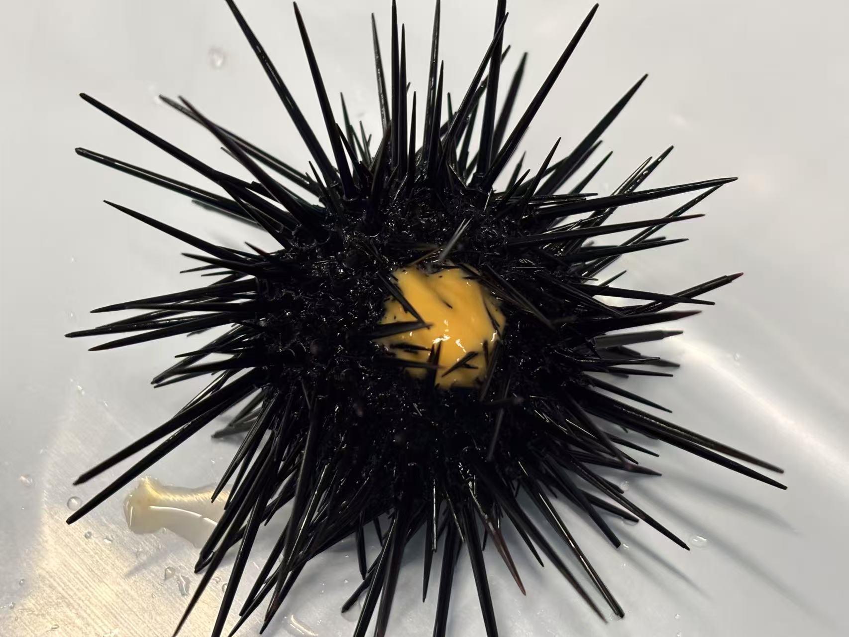
Figure 1. Adult sea urchins releasing gametes following KCl injection.
Egg collection and washing
Eggs were gently collected using wide-bore pipettes to avoid mechanical damage.
To remove debris and excess coelomic fluid, eggs were washed three times with filtered seawater:
- Eggs allowed to settle by gravity
- Supernatant carefully removed
- Fresh filtered seawater added
This ensured high-quality eggs for fertilization.
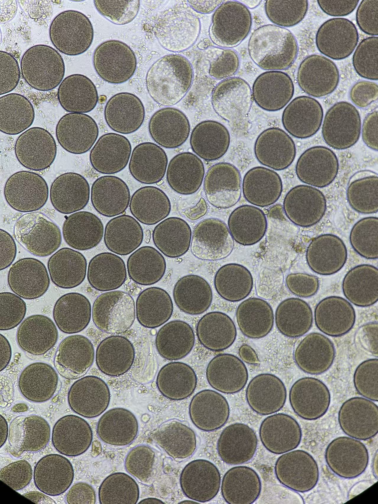
Figure 2. Collected eggs after washing in filtered seawater.
Fertilization
Concentrated sperm was diluted in seawater to create a working sperm solution.
A small volume of diluted sperm was added dropwise to the egg suspension while gently swirling.
Successful fertilization was confirmed by the appearance of a fertilization membrane around eggs within several minutes.
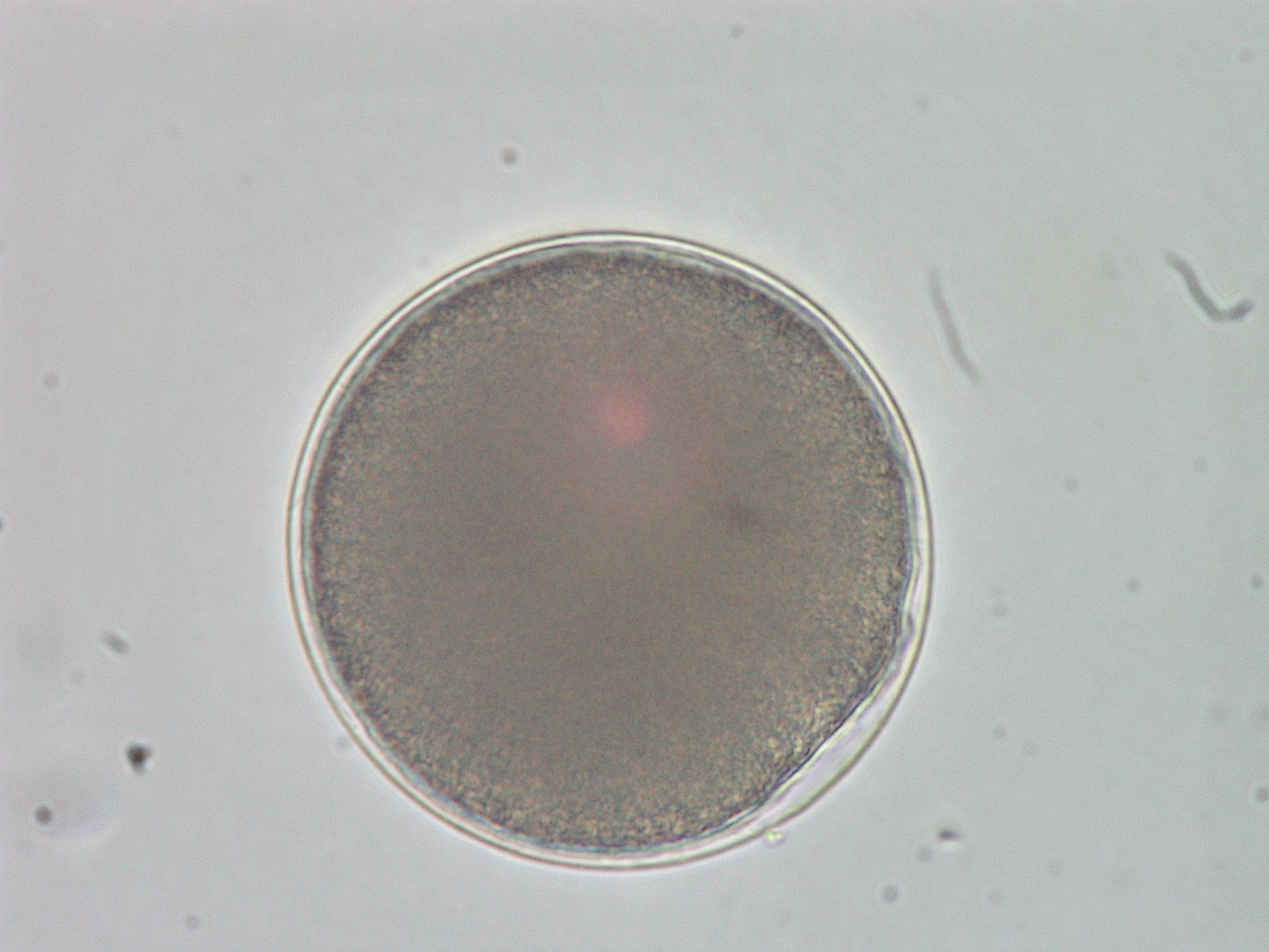
Figure 3. Fertilized eggs with visible fertilization envelopes.
Early embryonic development
Following fertilization, embryos were maintained in gently aerated culture vessels under defined:
- Temperature treatments
- Salinity treatments
Cleavage occurred rapidly and synchronously, with embryos progressing through a series of well-defined developmental stages from early cell division to gastrulation (Figure 4).
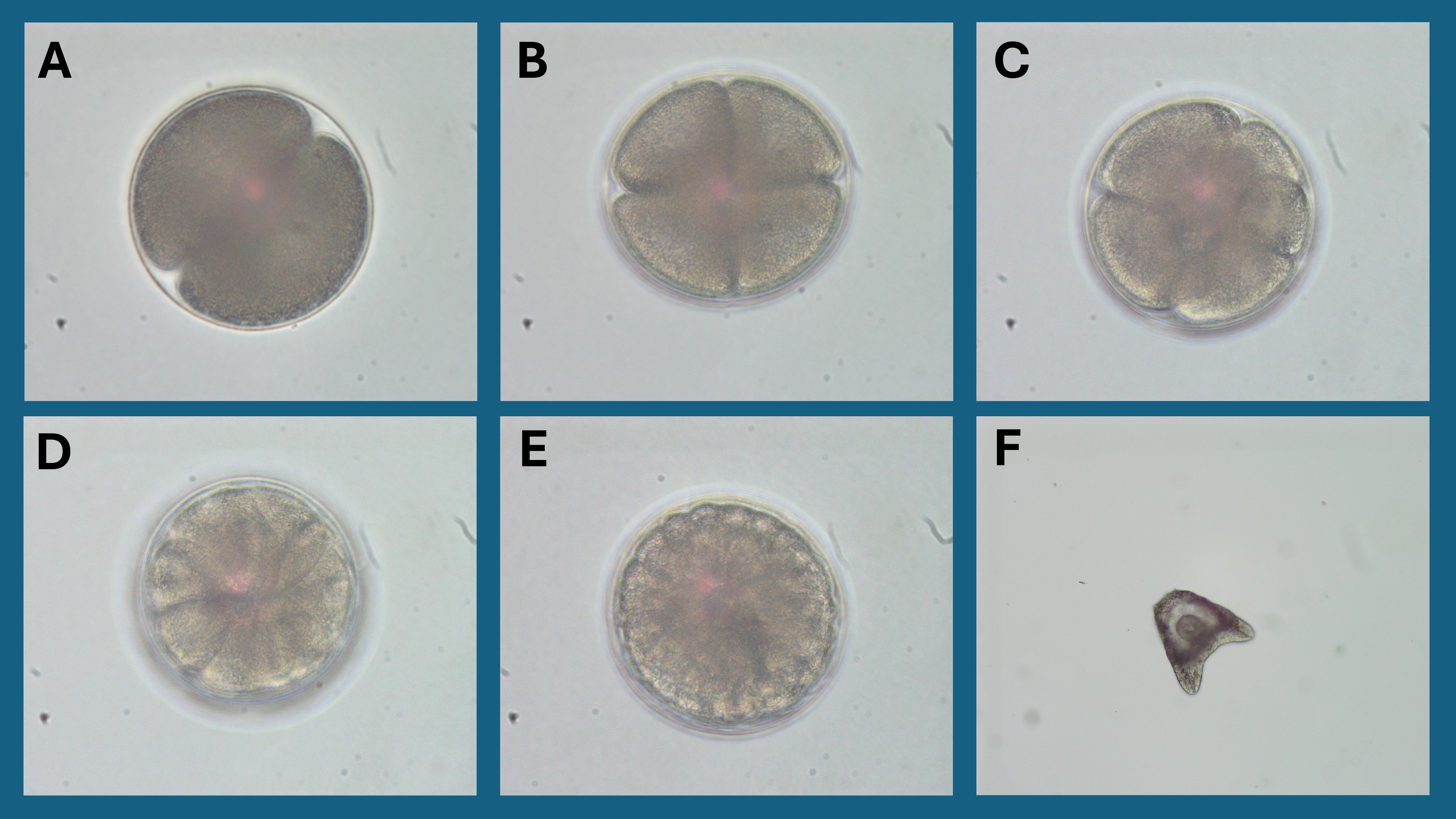
Figure 4. Early embryonic development stages. (A) Two-cell stage shortly after the first cleavage division, showing two equal-sized blastomeres within the fertilization envelope. (B) Four-cell stage following the second cleavage, with blastomeres arranged symmetrically. (C) Eight-cell stage as continued cleavage increases cell number and reduces individual cell size. (D) Sixteen-cell stage, marking the transition toward more compact cellular organization. (E) Blastula stage, characterized by a spherical embryo with a developing blastocoel and a smooth outer cell layer. (F) Gastrula stage, showing clear morphological differentiation and the onset of gastrulation with tissue invagination.
Two-arm larval stage
After several days, larvae developed characteristic skeletal arms, marking the planktonic feeding stage.
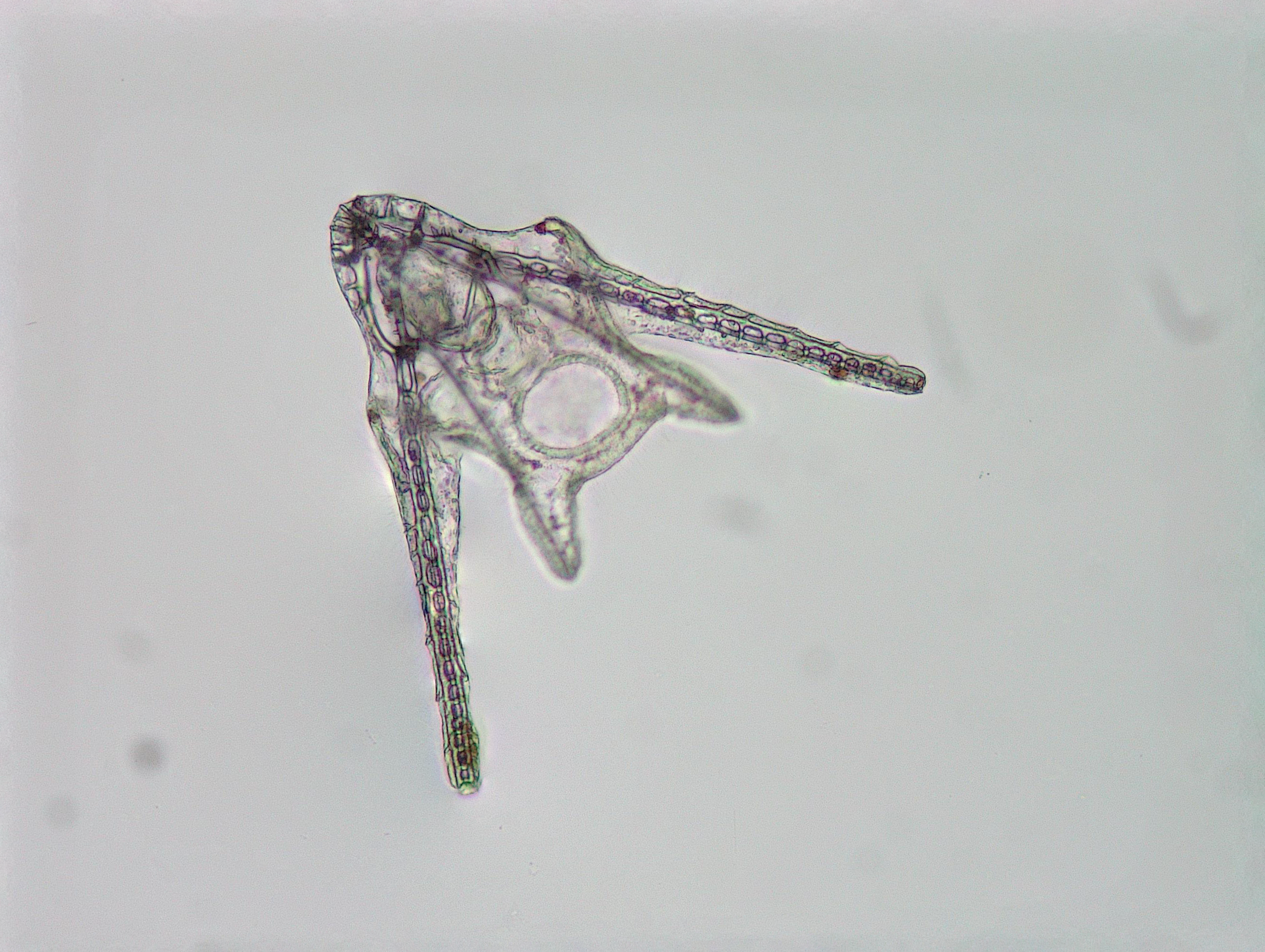
Figure 5. Two-arm pluteus larvae during early development.
Six-arm larval stage
With continued growth and feeding, larvae extended additional arms and increased in size.
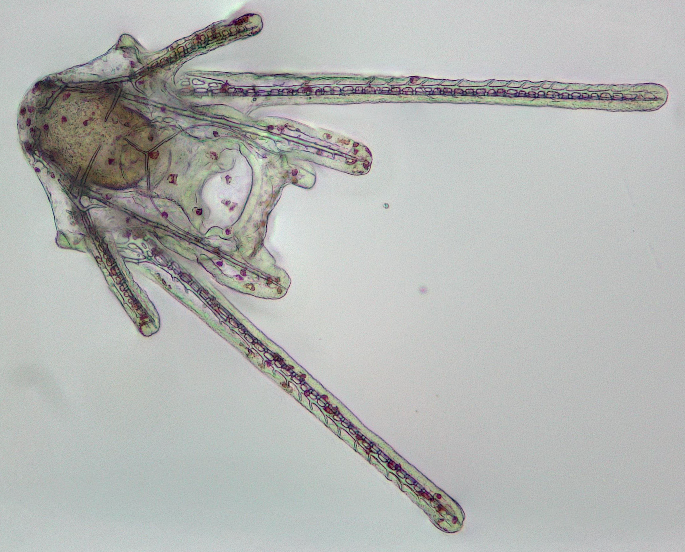
Figure 6. Six-arm larvae showing advanced skeletal development.
Metamorphosis
Settlement and metamorphic transition
Competent larvae eventually settled onto suitable surfaces and initiated metamorphosis.
This process involved:
- Loss of larval arms
- Formation of radial symmetry
- Development of juvenile structures
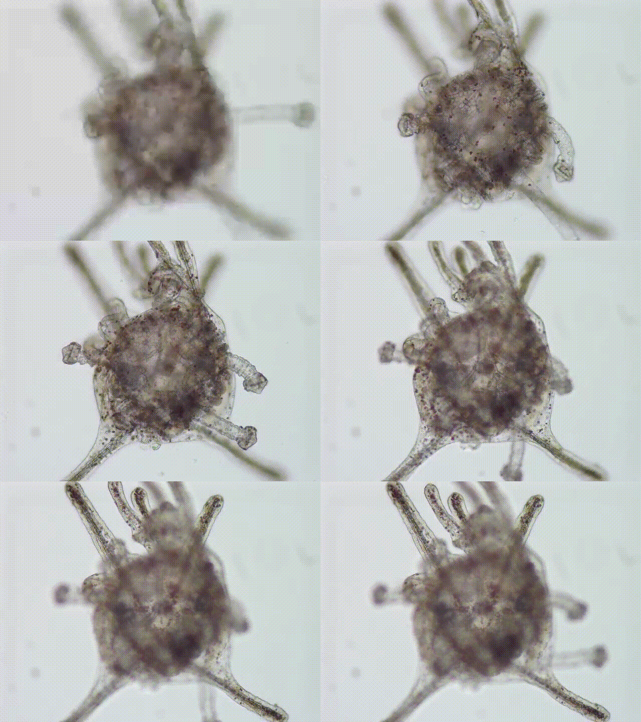
Figure 7. Metamorphic transition from larva to juvenile.
Abnormal metamorphosis under thermal and salinity stress
Under combined temperature and salinity treatments, a subset of larvae exhibited abnormal or incomplete metamorphosis (Figure 8). These individuals showed disrupted morphological transitions, including irregular body shapes, partial reabsorption of larval structures, and malformed juvenile features.
Such abnormalities suggest that environmental stress during early development can interfere with the tightly regulated processes of settlement and tissue reorganization, potentially reducing post-settlement survival and recruitment success.
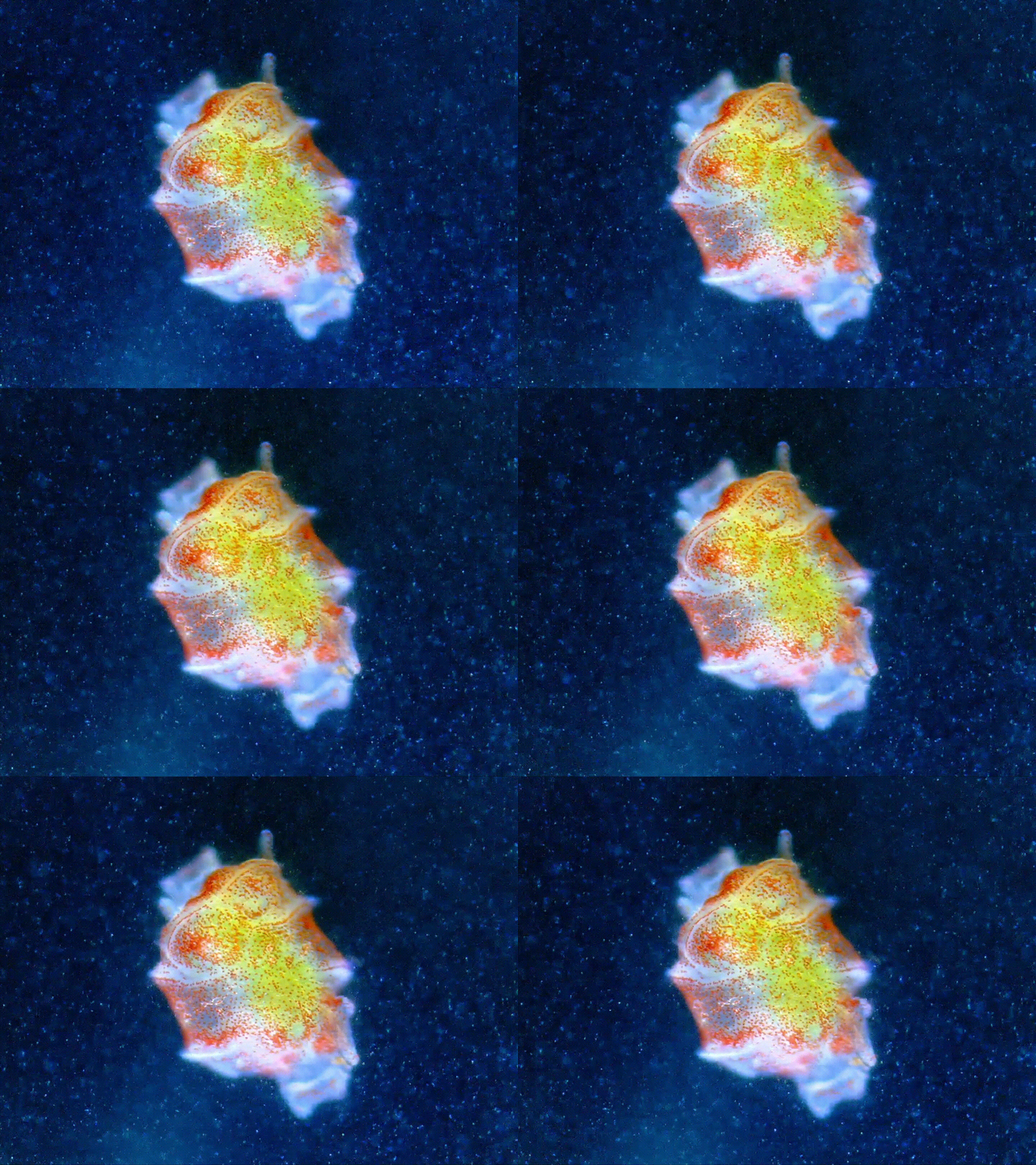
Figure 8. Abnormal metamorphic development under combined temperature and salinity treatments. Larvae exposed to elevated temperature and altered salinity showed incomplete or abnormal metamorphic transitions. In this image, two individuals appear to be partially fused, suggesting strong developmental disturbance under stress conditions.
Juvenile sea urchins
Following metamorphosis, individuals developed into benthic juveniles with visible spines and test structure.
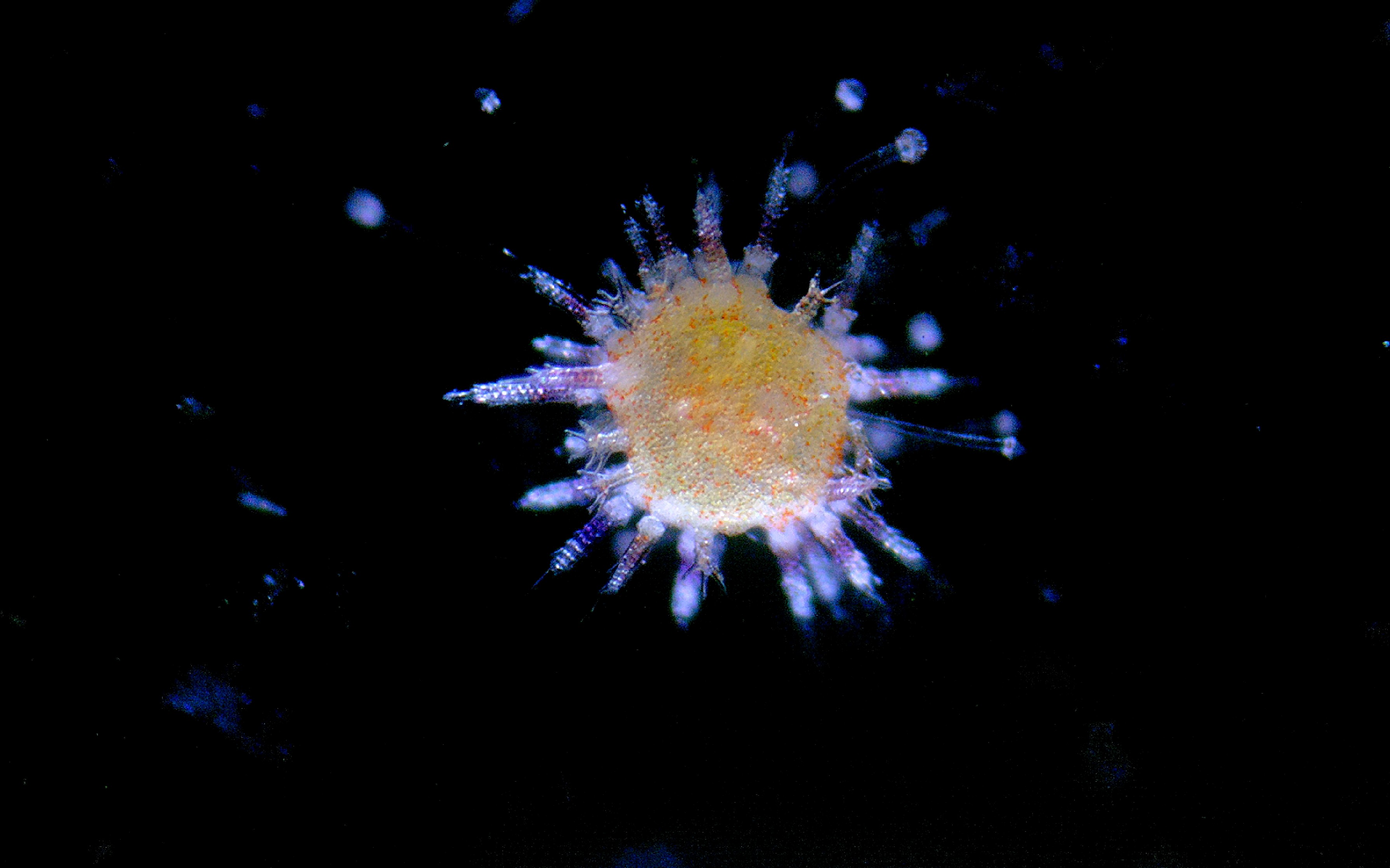
Figure 9. Newly formed juvenile sea urchins.
Experimental significance
Tracking individuals from fertilization through juvenile stages allows assessment of:
- Developmental sensitivity to temperature elevation
- Impacts of salinity stress on growth and survival
- Carry-over effects across life stages
These early-life responses play a crucial role in shaping population resilience under increasing climate extremes.
Final reflections
Culturing sea urchins across complete developmental cycles provides direct insight into how environmental stress influences:
- Developmental timing
- Morphological integrity
- Settlement success
The images presented here reflect both the technical process and the biological consequences of climate-driven stressors.
END
Copyright notice:
© 2026 Zhengquan Zhou. All images and figures presented in this blog are the intellectual property of the author and are protected by copyright. Unauthorized reproduction or distribution is prohibited without written permission. Academic and educational use is permitted with proper citation of this page.